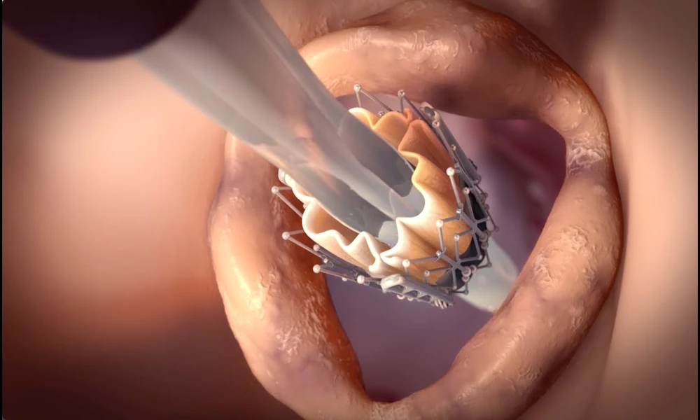
TAVR, also called TAVI, provides a catheter-based way to replace a severely narrowed aortic valve. For suitable patients, it can avoid the conventional open-chest approach of surgical valve replacement and may allow a shorter physical recovery. That does not make it a risk-free procedure.
The safest way to think about TAVR is not to ask whether complications are possible, because every major cardiac intervention carries risk. The more useful question is how an experienced team identifies a patient’s specific risks before treatment and plans the procedure to reduce them.
Why Patient Evaluation Comes Before Procedure Selection
Severe aortic stenosis does not automatically mean that TAVR is the best option. The Heart Team first confirms the diagnosis, reviews symptoms and compares transcatheter treatment with surgical valve replacement.
Age, life expectancy, frailty, previous heart surgery, kidney function, lung disease, coronary disease, other valve problems and the patient’s long-term treatment pathway all matter.
A careful TAVI procedure evaluation also examines anatomy in detail. The valve itself may be suitable, but small or heavily calcified access arteries, low coronary arteries or unusual aortic-root anatomy can change procedural risk.
Vascular Bleeding and Access-Site Complications
Most TAVR procedures are performed through the femoral artery in the groin. Because relatively large catheters must pass through the artery, bleeding, vessel injury, blockage or dissection can occur.
CT imaging before the procedure helps measure the arteries and identify calcification or narrowing. If the femoral route appears unsafe, the team may consider another strategy.
During and after the procedure, the access site is monitored carefully. Experienced teams also need the ability to treat vascular complications rapidly if they occur.
Stroke Risk
Small particles of calcium, tissue or clot can occasionally travel through the bloodstream during valve manipulation and reach the brain, causing a stroke.
The overall risk is influenced by the patient’s age, vascular disease, aortic anatomy and other clinical factors. The procedural team uses careful catheter technique and anticoagulation strategies to reduce this risk.
Some patients may be considered for cerebral embolic protection devices, although their use depends on individual circumstances and local practice.
Heart Rhythm and Pacemaker Risk
The aortic valve lies close to the heart’s electrical conduction system. When the new valve frame expands, it can affect nearby conduction tissue.
Some patients develop new bundle-branch block or heart block after TAVR. Rhythm changes may resolve, but a proportion of patients require a permanent pacemaker.
An ECG before treatment establishes the baseline rhythm. Valve type, implant depth and pre-existing electrical abnormalities can influence risk, so monitoring continues after the procedure.
Leakage Around the New Valve
Because a transcatheter valve is implanted inside the calcified native valve rather than after surgically removing it, a small amount of blood can sometimes leak around the outside of the replacement valve. This is called paravalvular regurgitation.
Modern valve designs and better CT-based sizing have reduced significant leakage, but the team still checks for it immediately after implantation with imaging and haemodynamic measurements.
If leakage is important, additional treatment may sometimes be required during the same procedure.
Kidney Injury
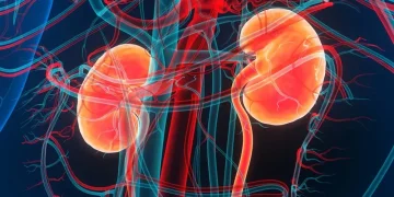
Contrast dye is commonly used during CT planning and the procedure. Older adults with aortic stenosis may already have reduced kidney function, making this an important consideration.
A pre-operative assessment reviews creatinine and other kidney measurements. The team may adjust hydration, minimise contrast exposure and review medications that could affect kidney function.
Kidney risk is another reason TAVR preparation should be personalised rather than based on a fixed checklist.
Coronary Obstruction
The coronary arteries arise close to the aortic valve. In certain anatomies, displaced native valve tissue can interfere with blood flow into a coronary artery when the replacement valve is deployed.
CT planning helps identify low coronary heights, narrow sinuses and other anatomical features associated with higher risk.
When risk is identified, specialised protective or alternative strategies may be considered.
Valve Positioning and Annular Injury
Accurate positioning is essential. A valve placed too high or too low may not function as intended and can affect surrounding structures.
Heavy calcium and certain anatomical patterns may also increase the risk of injury to the aortic annulus or root. Detailed imaging, appropriate device sizing and controlled deployment are central to avoiding these complications.
Why a Pre-Operative Assessment Matters
A thorough pre-operative assessment does more than identify a list of possible problems. It allows the Heart Team to connect each risk to a prevention or management strategy.
Echocardiography confirms valve severity. CT maps the valve and access arteries. Coronary assessment identifies blockages. ECG shows baseline conduction. Blood tests evaluate kidney function, anaemia and clotting. Clinical review identifies frailty and other illnesses.
Patients can review the pre-operative assessment before TAVR for more detail.
Risk Does Not Mean the Procedure Should Be Avoided
Patients sometimes become alarmed when they hear a detailed complication list. The purpose of discussing risk is not to discourage treatment. Severe symptomatic aortic stenosis itself can carry significant consequences if appropriate valve replacement is delayed.
The decision therefore compares the risks of TAVR, the risks of surgery and the risks of leaving the valve untreated.
What Happens After the Procedure?
Close monitoring continues after implantation because some complications, particularly rhythm problems, vascular bleeding or kidney changes, may become evident during the first hours or days.
Good post-procedure TAVI care includes clinical assessment, ECG monitoring, blood tests, access-site review and echocardiography. Clear discharge instructions help patients recognise warning signs after returning home.
The Bottom Line
TAVR is a heart-valve procedure performed through a less invasive route. Its safety depends not only on technical skill during valve deployment but also on the quality of evaluation that comes before it.
Detailed imaging, medical assessment and Heart Team discussion help identify vascular, neurological, rhythm, kidney and anatomical risks before treatment. That information allows the team to modify the plan, prepare for complications and decide whether TAVR or surgery is the better approach. Proper evaluation turns risk assessment into practical procedural planning rather than a simple list of possible complications.